

Which Of The Following Statements Regarding The Cell Cycle Control System Is False?
Chapter 6: Introduction to Reproduction at the Cellular Level
6.2 The Cell Cycle
Learning Objectives
By the stop of this department, you will be able to:
- Draw the three stages of interphase
- Discuss the behavior of chromosomes during mitosis and how the cytoplasmic content divides during cytokinesis
- Ascertain the quiescent 10000 stage
- Explicate how the three internal control checkpoints occur at the terminate of G1, at the Thou2–M transition, and during metaphase
The jail cell cycle is an ordered series of events involving prison cell growth and prison cell sectionalisation that produces two new daughter cells. Cells on the path to cell division proceed through a series of precisely timed and carefully regulated stages of growth, Dna replication, and sectionalisation that produce two genetically identical cells. The cell cycle has two major phases: interphase and the mitotic phase (Effigy 6.3). During interphase, the cell grows and DNA is replicated. During the mitotic phase, the replicated DNA and cytoplasmic contents are separated and the cell divides.
Picket this video about the cell wheel: https://www.youtube.com/watch?five=Wy3N5NCZBHQ
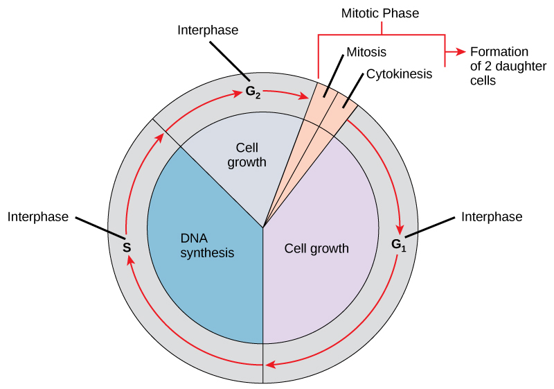
Interphase
During interphase, the cell undergoes normal processes while too preparing for jail cell partitioning. For a prison cell to move from interphase to the mitotic stage, many internal and external conditions must be met. The 3 stages of interphase are called Chiliad1, S, and G2.
G1 Phase
The first stage of interphase is called the Gone stage, or first gap, because picayune change is visible. However, during the Grand1 stage, the jail cell is quite active at the biochemical level. The prison cell is accumulating the edifice blocks of chromosomal DNA and the associated proteins, equally well as accumulating enough energy reserves to consummate the task of replicating each chromosome in the nucleus.
S Phase
Throughout interphase, nuclear Dna remains in a semi-condensed chromatin configuration. In the S stage (synthesis stage), Dna replication results in the germination of two identical copies of each chromosome—sister chromatids—that are firmly attached at the centromere region. At this stage, each chromosome is made of two sister chromatids and is a duplicated chromosome. The centrosome is duplicated during the Due south phase. The 2 centrosomes will give rising to the mitotic spindle, the apparatus that orchestrates the move of chromosomes during mitosis. The centrosome consists of a pair of rod-like centrioles at right angles to each other. Centrioles help organize jail cell partition. Centrioles are not present in the centrosomes of many eukaryotic species, such equally plants and most fungi.
M2 Phase
In the Chiliad2 stage, or second gap, the cell replenishes its energy stores and synthesizes the proteins necessary for chromosome manipulation. Some cell organelles are duplicated, and the cytoskeleton is dismantled to provide resources for the mitotic spindle. There may be additional jail cell growth during One thousand2. The terminal preparations for the mitotic stage must be completed before the cell is able to enter the starting time stage of mitosis.
The Mitotic Phase
To brand two daughter cells, the contents of the nucleus and the cytoplasm must be divided. The mitotic phase is a multistep process during which the duplicated chromosomes are aligned, separated, and moved to opposite poles of the cell, and then the cell is divided into 2 new identical daughter cells. The first portion of the mitotic phase, mitosis, is composed of five stages, which accomplish nuclear division. The 2d portion of the mitotic phase, chosen cytokinesis, is the physical separation of the cytoplasmic components into 2 daughter cells.
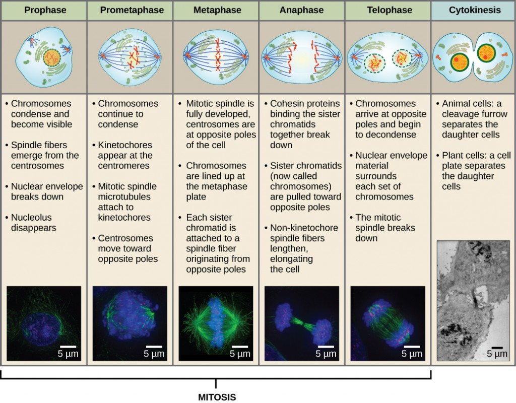
Mitosis
Mitosis is divided into a series of phases—prophase, prometaphase, metaphase, anaphase, and telophase—that result in the division of the cell nucleus (Figure half dozen.4).
Which of the post-obit is the correct gild of events in mitosis?
- Sister chromatids line upwards at the metaphase plate. The kinetochore becomes attached to the mitotic spindle. The nucleus re-forms and the jail cell divides. The sister chromatids separate.
- The kinetochore becomes attached to the mitotic spindle. The sister chromatids split up. Sister chromatids line up at the metaphase plate. The nucleus re-forms and the jail cell divides.
- The kinetochore becomes attached to metaphase plate. Sister chromatids line up at the metaphase plate. The kinetochore breaks downwardly and the sister chromatids separate. The nucleus re-forms and the prison cell divides.
- The kinetochore becomes fastened to the mitotic spindle. Sister chromatids line up at the metaphase plate. The kinetochore breaks apart and the sis chromatids separate. The nucleus re-forms and the cell divides.
During prophase, the "first phase," several events must occur to provide admission to the chromosomes in the nucleus. The nuclear envelope starts to intermission into modest vesicles, and the Golgi appliance and endoplasmic reticulum fragment and disperse to the periphery of the jail cell. The nucleolus disappears. The centrosomes begin to move to contrary poles of the prison cell. The microtubules that form the ground of the mitotic spindle extend between the centrosomes, pushing them farther autonomously as the microtubule fibers lengthen. The sister chromatids begin to coil more tightly and become visible under a light microscope.
During prometaphase, many processes that were begun in prophase keep to advance and culminate in the formation of a connectedness betwixt the chromosomes and cytoskeleton. The remnants of the nuclear envelope disappear. The mitotic spindle continues to develop equally more microtubules gather and stretch across the length of the former nuclear area. Chromosomes go more condensed and visually detached. Each sister chromatid attaches to spindle microtubules at the centromere via a protein circuitous chosen the kinetochore.
During metaphase, all of the chromosomes are aligned in a plane called the metaphase plate, or the equatorial plane, midway between the 2 poles of the prison cell. The sister chromatids are however tightly fastened to each other. At this fourth dimension, the chromosomes are maximally condensed.
During anaphase, the sister chromatids at the equatorial airplane are carve up autonomously at the centromere. Each chromatid, now chosen a chromosome, is pulled chop-chop toward the centrosome to which its microtubule was fastened. The cell becomes visibly elongated every bit the non-kinetochore microtubules slide confronting each other at the metaphase plate where they overlap.
During telophase, all of the events that prepare up the duplicated chromosomes for mitosis during the first three phases are reversed. The chromosomes reach the opposite poles and begin to decondense (unravel). The mitotic spindles are broken down into monomers that volition be used to gather cytoskeleton components for each girl cell. Nuclear envelopes form around chromosomes.
Concept in Activeness

This page of movies illustrates different aspects of mitosis. Watch the movie entitled "DIC microscopy of prison cell division in a newt lung cell" and identify the phases of mitosis.
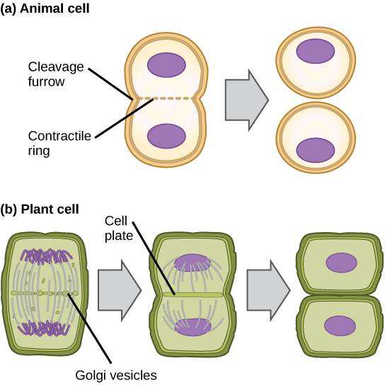
Cytokinesis
Cytokinesis is the second part of the mitotic phase during which cell partitioning is completed past the physical separation of the cytoplasmic components into 2 girl cells. Although the stages of mitosis are like for most eukaryotes, the process of cytokinesis is quite dissimilar for eukaryotes that accept cell walls, such as plant cells.
In cells such every bit animal cells that lack prison cell walls, cytokinesis begins following the onset of anaphase. A contractile band equanimous of actin filaments forms merely inside the plasma membrane at the former metaphase plate. The actin filaments pull the equator of the jail cell inward, forming a cleft. This fissure, or "fissure," is called the cleavage furrow. The furrow deepens as the actin ring contracts, and somewhen the membrane and cell are broken in ii (Effigy 6.5).
In establish cells, a cleavage furrow is not possible because of the rigid jail cell walls surrounding the plasma membrane. A new jail cell wall must form between the girl cells. During interphase, the Golgi apparatus accumulates enzymes, structural proteins, and glucose molecules prior to breaking upwards into vesicles and dispersing throughout the dividing jail cell. During telophase, these Golgi vesicles motion on microtubules to collect at the metaphase plate. At that place, the vesicles fuse from the centre toward the cell walls; this construction is called a cell plate. Equally more than vesicles fuse, the cell plate enlarges until it merges with the cell wall at the periphery of the prison cell. Enzymes employ the glucose that has accumulated between the membrane layers to build a new cell wall of cellulose. The Golgi membranes become the plasma membrane on either side of the new cell wall (Figure vi.5).
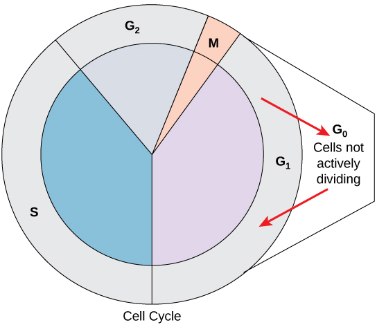
Yard0 Phase
Not all cells adhere to the classic jail cell-cycle pattern in which a newly formed daughter cell immediately enters interphase, closely followed by the mitotic phase. Cells in the M0 phase are not actively preparing to divide. The cell is in a quiescent (inactive) stage, having exited the jail cell cycle. Some cells enter Yard0 temporarily until an external betoken triggers the onset of 10001. Other cells that never or rarely split, such equally mature cardiac muscle and nerve cells, remain in Thou0 permanently (Figure 6.half-dozen).
Command of the Cell Wheel
The length of the cell cycle is highly variable even within the cells of an individual organism. In humans, the frequency of cell turnover ranges from a few hours in early embryonic evolution to an average of two to five days for epithelial cells, or to an entire homo lifetime spent in G0 past specialized cells such as cortical neurons or cardiac muscle cells. There is besides variation in the fourth dimension that a cell spends in each phase of the cell cycle. When fast-dividing mammalian cells are grown in civilisation (outside the body under optimal growing weather), the length of the bicycle is approximately 24 hours. In rapidly dividing man cells with a 24-hour cell bike, the G1 phase lasts approximately 11 hours. The timing of events in the cell bike is controlled by mechanisms that are both internal and external to the prison cell.
Regulation at Internal Checkpoints
It is essential that daughter cells be exact duplicates of the parent cell. Mistakes in the duplication or distribution of the chromosomes lead to mutations that may be passed forrad to every new prison cell produced from the abnormal cell. To forbid a compromised cell from continuing to separate, there are internal control mechanisms that operate at three main cell cycle checkpoints at which the cell cycle tin can exist stopped until weather condition are favorable. These checkpoints occur near the end of Chiliadone, at the Yardtwo–Thou transition, and during metaphase (Figure half-dozen.7).
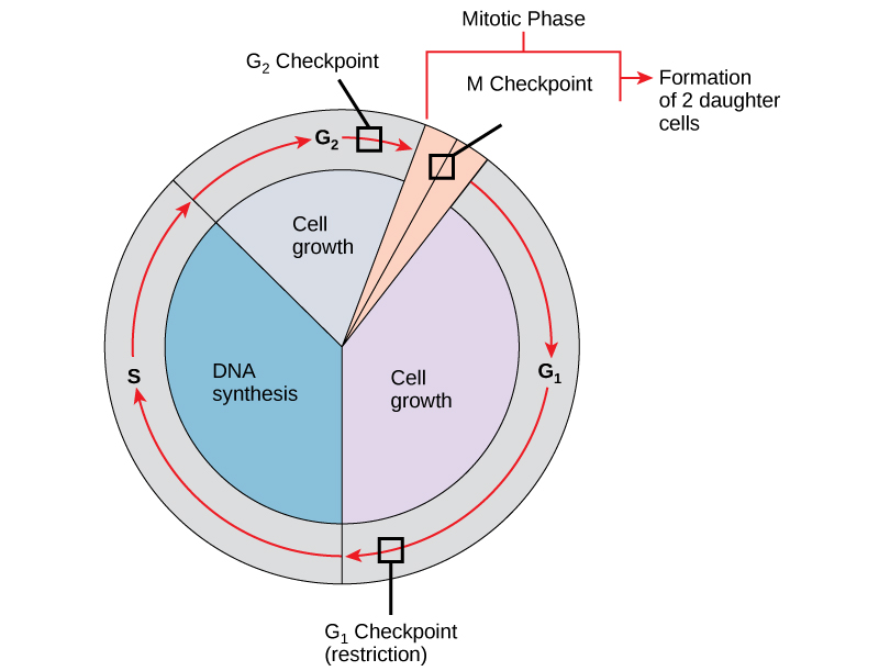
The G1 Checkpoint
The Yard1 checkpoint determines whether all atmospheric condition are favorable for cell sectionalization to keep. The Gone checkpoint, also chosen the restriction bespeak, is the point at which the cell irreversibly commits to the prison cell-division process. In addition to acceptable reserves and cell size, there is a check for damage to the genomic Deoxyribonucleic acid at the Chiliad1 checkpoint. A cell that does not meet all the requirements volition not be released into the Due south phase.
The Gii Checkpoint
The Grandii checkpoint confined the entry to the mitotic phase if certain conditions are non met. Equally in the Gi checkpoint, jail cell size and protein reserves are assessed. Even so, the near of import role of the Thousandtwo checkpoint is to ensure that all of the chromosomes have been replicated and that the replicated DNA is not damaged.
The K Checkpoint
The M checkpoint occurs near the end of the metaphase stage of mitosis. The M checkpoint is also known equally the spindle checkpoint because it determines if all the sister chromatids are correctly attached to the spindle microtubules. Because the separation of the sister chromatids during anaphase is an irreversible step, the cycle will not proceed until the kinetochores of each pair of sis chromatids are firmly anchored to spindle fibers arising from opposite poles of the prison cell.
Concept in Activeness

Watch what occurs at the Grand1, 10002, and Grand checkpoints by visiting this animation of the jail cell cycle.
Section Summary
The cell bicycle is an orderly sequence of events. Cells on the path to prison cell division proceed through a series of precisely timed and carefully regulated stages. In eukaryotes, the cell wheel consists of a long preparatory menstruum, called interphase. Interphase is divided into Kone, S, and G2 phases. Mitosis consists of five stages: prophase, prometaphase, metaphase, anaphase, and telophase. Mitosis is unremarkably accompanied by cytokinesis, during which the cytoplasmic components of the daughter cells are separated either by an actin ring (animal cells) or past cell plate formation (plant cells).
Each footstep of the cell cycle is monitored past internal controls called checkpoints. At that place are 3 major checkpoints in the cell cycle: i about the end of Gane, a second at the Yard2–M transition, and the third during metaphase.
Glossary
anaphase : the stage of mitosis during which sis chromatids are separated from each other
cell cycle : the ordered sequence of events that a cell passes through betwixt one cell partitioning and the next
cell wheel checkpoints: mechanisms that monitor the preparedness of a eukaryotic cell to advance through the various cell wheel stages
cell plate: a structure formed during establish-cell cytokinesis past Golgi vesicles fusing at the metaphase plate; will ultimately lead to germination of a prison cell wall to dissever the two daughter cells
centriole: a paired rod-similar structure constructed of microtubules at the center of each animate being cell centrosome
cleavage furrow: a constriction formed by the actin band during beast-cell cytokinesis that leads to cytoplasmic sectionalisation
cytokinesis: the sectionalization of the cytoplasm following mitosis to grade two daughter cells
Thousand0 phase: a jail cell-cycle stage distinct from the One thousandone phase of interphase; a jail cell in Grand0 is not preparing to divide
Chiliadone phase : (as well, showtime gap) a jail cell-cycle stage; first stage of interphase centered on cell growth during mitosis
G2 stage: (also, second gap) a cell-cycle phase; tertiary phase of interphase where the cell undergoes the terminal preparations for mitosis
interphase: the period of the cell wheel leading up to mitosis; includes Chiliadone, S, and G2 phases; the acting between two sequent cell divisions
kinetochore: a protein structure in the centromere of each sister chromatid that attracts and binds spindle microtubules during prometaphase
metaphase plate: the equatorial airplane midway betwixt two poles of a cell where the chromosomes align during metaphase
metaphase : the stage of mitosis during which chromosomes are lined up at the metaphase plate
mitosis: the period of the jail cell wheel at which the duplicated chromosomes are separated into identical nuclei; includes prophase, prometaphase, metaphase, anaphase, and telophase
mitotic phase: the period of the cell wheel when duplicated chromosomes are distributed into ii nuclei and the cytoplasmic contents are divided; includes mitosis and cytokinesis
mitotic spindle: the microtubule apparatus that orchestrates the motility of chromosomes during mitosis
prometaphase : the stage of mitosis during which mitotic spindle fibers attach to kinetochores
prophase: the phase of mitosis during which chromosomes condense and the mitotic spindle begins to grade
quiescent: describes a prison cell that is performing normal prison cell functions and has not initiated preparations for cell division
S stage: the second, or synthesis phase, of interphase during which DNA replication occurs
telophase: the phase of mitosis during which chromosomes arrive at opposite poles, decondense, and are surrounded by new nuclear envelopes

Which Of The Following Statements Regarding The Cell Cycle Control System Is False?,
Source: https://opentextbc.ca/biology/chapter/6-2-the-cell-cycle/
Posted by: tarverfrose1966.blogspot.com
0 Response to "Which Of The Following Statements Regarding The Cell Cycle Control System Is False?"
Post a Comment